Bones in health and archaeology
The skeleton is essential for human health, but also an exciting hierarchical composite material. Our understanding of both bone biology and how structure and mechanical functions are connected remains disappointingly incomplete. This is in particular due to the lack of multiscale 3D understanding and/or techniques. This problem is a major stumbling block in our understanding of musculoskeletal disease, development of efficient treatments and diagnostics, e.g. within osteoporosis. In archaeology, bones are of special interest since they are often all that remains. Bones are therefore a potential treasure trove of insights into how our ancestors lived, if only we understood how bones change upon environmental exposure during the many years for example underground (diagenesis). Other hard biological tissues, e.g. shells, eggs, pearls, teeth etc. are also of great interest from both a materials and archaeological perspective.
The tools of SOLID will thus be directly relevant for both biological, medical, forensic and archaeological research. The developments in X-ray and neutron imaging will be game changers within these areas due to novel imaging contrast mechanisms and X-ray imaging with nanoscale resolution can give insights into interactions between bones, the cells of the body and musculoskeletal diseases. One central challenge is the fact that bone is a living material traversed by liquids such as blood. One SOLID goal is to study liquid transport by neutron imaging to change our understanding of bone from a static skeleton to the living, dynamical tissue it really is. This is important for bone mechanics but also for bone repair (remodeling) and for efficient delivery of drugs.
The tools of SOLID will thus be directly relevant for both biological, medical, forensic and archaeological research. The developments in X-ray and neutron imaging will be game changers within these areas due to novel imaging contrast mechanisms and X-ray imaging with nanoscale resolution can give insights into interactions between bones, the cells of the body and musculoskeletal diseases. One central challenge is the fact that bone is a living material traversed by liquids such as blood. One SOLID goal is to study liquid transport by neutron imaging to change our understanding of bone from a static skeleton to the living, dynamical tissue it really is. This is important for bone mechanics but also for bone repair (remodeling) and for efficient delivery of drugs.
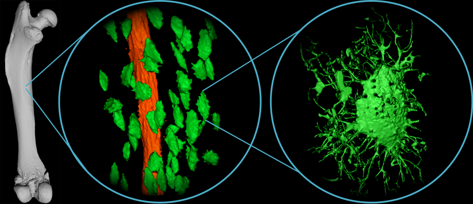
The upper Figure is part of an article in the Danish journal Aktuel Naturvidenskab called 3D X-ray reveals the inside of the bones (in Danish: 3D-Røntgensyn afslører knoglernes indre), by Nina Wittig and Henrik Birkedal.